January 13th 2026
Understanding the Role of Neuronavigation in TMS
Transcranial Magnetic Stimulation (TMS) is a well-established, non-invasive technique used in both clinical practice and neuroscience research. Over time, attention has expanded beyond stimulation protocols alone to include how stimulation targets are identified, applied, and reproduced across sessions.
One approach that has gained increasing attention in this context is neuronavigation.
Neuronavigation is not a requirement for delivering TMS. However, it has become a relevant tool for clinicians and researchers who prioritize precision, reproducibility, and structured documentation in their workflows.
What Is Neuronavigation in TMS?
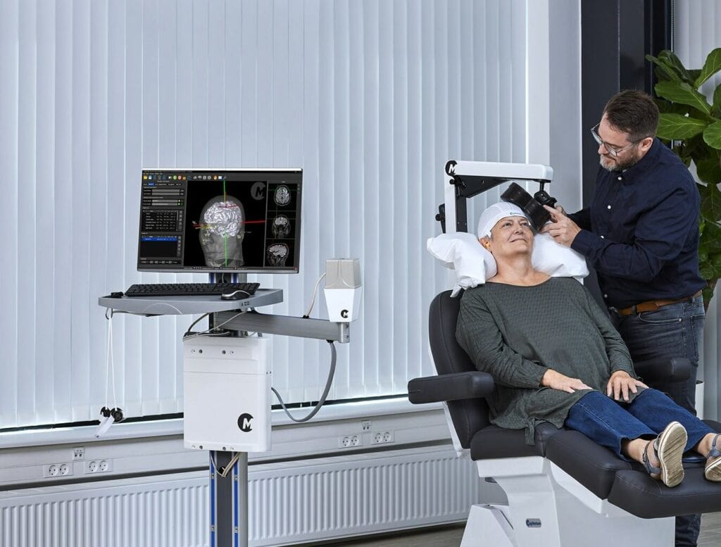
Neuronavigation refers to systems that link TMS coil positioning to brain anatomy, enabling visualization and tracking of the stimulation target in real time. Depending on the setup, navigation may be based on standardized anatomical landmarks, template-based brain models, or individual structural imaging data where available1,2.
By providing visual feedback on coil position and orientation1, neuronavigation supports a more transparent and consistent approach to TMS targeting3.
Why Precision and Reproducibility Matter
In traditional TMS workflows, targeting is often guided by external landmarks and operator experience. While this approach is widely used, it can introduce variability—particularly across multi-session treatment courses, different operators, or longitudinal research protocols1.
Neuronavigation helps reduce this variability by supporting consistent coil placement and enabling verification of stimulation location over time1,4. For many users, this contributes to greater confidence in daily workflows and study design.
Applications in Clinical and Research Settings
Neuronavigation can add value across a range of professional environments.
Clinical Settings
In clinical practice, neuronavigation may support standardized workflows by helping clinicians maintain consistent targeting even when staff rotate or patient positioning varies between sessions⁴. Visual representation of the stimulation target can also support communication when discussing treatment planning with patients.
Research Settings
In research environments, neuronavigation is often used to support documentation, reproducibility, and alignment with imaging-based targeting approaches. These elements are particularly relevant for advanced study designs, longitudinal data collection, and publication requirements.
Across both settings, neuronavigation provides a structured method for linking stimulation delivery to anatomical reference points1.
Supporting Workflow Confidence
Beyond technical precision, neuronavigation may also contribute to workflow confidence. By reducing reliance on manual estimation alone, navigation tools allow clinicians and researchers to verify that stimulation is delivered as intended1.
This transparency can be particularly relevant in multi-session protocols, multi-operator environments, and long-term studies.
Clear and Responsible Communication
As neuronavigation becomes more visible in TMS practice, clear and accurate communication is essential. Neuronavigation should be understood as a supportive tool—one that enhances precision and reproducibility—rather than a requirement or a guarantee of clinical outcomes1.
Maintaining this distinction helps ensure aligned expectations among clinicians, researchers, partners, and patients, and supports responsible use of the technology.
Looking Ahead
Neuronavigation continues to play a role in how TMS workflows evolve. By supporting accurate targeting, reproducibility, and structured documentation, neuronavigation may help clinics and research centers prepare for future developments while maintaining reliable stimulation delivery today4.
References
- Caulfield, K. A. et al. Neuronavigation maximizes accuracy and precision in TMS positioning: Evidence from 11,230 distance, angle, and electric field modeling measurements. Brain Stimulation 15, 1192–1205 (2022). https://doi.org/https://doi.org/10.1016/j.brs.2022.08.013
- Herwig, U., Satrapi, P. & Schonfeldt-Lecuona, C. Using the international 10-20 EEG system for positioning of transcranial magnetic stimulation. Brain Topogr 16, 95–99 (2003). https://doi.org/10.1023/b:brat.0000006333.93597.9d
- Lioumis, P. & Rosanova, M. The role of neuronavigation in TMS-EEG studies: Current applications and future perspectives. J Neurosci Methods 380, 109677 (2022). https://doi.org/10.1016/j.jneumeth.2022.109677
- Siebner, H. R. et al. Consensus paper: combining transcranial stimulation with neuroimaging. Brain Stimul 2, 58–80 (2009). https://doi.org/10.1016/j.brs.2008.11.002
Webinar Recording: How Neuronavigation is Transforming Clinical TMS
Watch this webinar recording for an engaging conversation on how neuronavigation is transforming the way clinicians deliver TMS.
In this webinar, Jón Gauti, Team Leader at The Brain Stimulation Clinic, Iceland, shares his hands-on experience of implementing a neuronavigation system in a real clinical setting.
Jón shares practical insights into:
- System integration
- Workflow optimization
- Day-to-day clinical use of neuronavigated TMS
What you’ll learn:
🔹 What neuronavigated TMS really means — and why it matters
🔹 Common misconceptions and practical benefits
🔹 Real user experiences and workflow integration
